 |
| Click to enlarge Patient's Resting ECG |
- 72 bpm
- Sinus rhythm
- Regular
- Normal
- PR - Normal (160ms)
- QRS - Normal (80ms)
- QT -360 ms (QTc ~395 ms Bazette)
- Prominence of T waves in anterior leads V2-4
- Small Q wave inferior leads
- Apparent pacing spike seen in leads V4-6 artefactual in nature
- Patient did not have a PPM in-situ
Interpretation:
- Essentially normal ECG
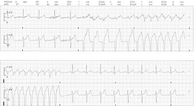
Rhythm Strip:
- Initial sinus rhythm, rate ~71bpm
- Multiple ventricular complexes
- Degeneration into broad complex tachycardia, rate ~175 bpm
- Spontaneous reversion to sinus rhythm
Interpretation:
- Episode of non-sustained VT
Serial troponin testing was normal but given captured VT and a period of chest / arm pain that preceded his Emergency Department attendance the patient was taken for angio, which showed:
- Mid LAD 70% stenosis
- LCx irregularities
- OM1 95% proximal stenosis
- RCA stent patnet but 30% re-stenosis
- PCI and stent to OM1 and mid LAD lesions
Post angio echo was normal and telemetry monitoring revealed no further episodes of VT, the VT episodes were felt to be ischaemic in origin.
References / Further Reading
Life in the Fast Lane
Textbook- Chan TC, Brady WJ, Harrigan RA, Ornato JP, Rosen P. ECG in Emergency Medicine and Acute Care. Elsevier Mosby 2005.

No comments:
Post a Comment