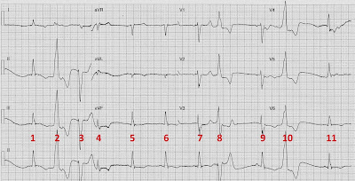
He was referred in to the Emergency Department by his GP having complained of nausea, lethargy, and anorexia for the preceding week. Past History of chronic renal failure,and ischaemic heart disease with previous 2 vessel PCI. Medications included frusemide, ACE-inhibitor, and a statin.
Check out the comments from our original post here.
I'll give you the punchline for this one up front as the patient presented with the following biochemistry results:
- Creatinine 421 umol/L [70-150]
- Patient's baseline creatinine ~300 umol/L
- Mg 0.17 mmol/L [0.65-1.10]
- Note K was normal
- Cor Cal 1.35 mmol/L [2.15-2.55]
- Ionised Ca 0.66 mmol/L [1.12 - 1.30]
- Phos 2.0 mmol/L [0.7-1.5]
- Alb 31 g/L [34-45]
 |
| Click to enlarge |
 |
| Number version of ECG Click to enlarge |
Rate:
- Mean ventricular rate ~66 bpm
- Sinus rhythm
- Complexes # 1, 5, 6, 7, 9, 11
- P waves difficult to see but best appreciated in leads V1 & V2
- Ventricular Ectopics
- Complexes # 2, 3, 8, 10
- Premature Junctional Complex
- Complex #4
- Normal
- PR - Normal (~130ms)
- QRS - Prolonged (140ms)
- QT - 490ms (QTc Bazette ~ 500 ms) - Prolonged
- Discordant ST / T wave changes with ventricular ectopic complexes
- T wave inversion sinus complexes leads I, II, II, aVL, V6
- Ventricular ectopics occur close to T wave especially complexes # 2 & 10
- Life-threatening electrolyte abnormalities
- ECG manifestations consistent with hypomagnesaemia and hypocalcaemia
- QT Prolongation
- QRS Prolongation
- Multiple ectopic complexes
- Risk of Torsades de Pointes
What happened?
The patient was admitted to the Critical Care Unit for cardiac monitoring and electrolyte replacement.
Further biochemistry revealed an elevated Parathyroid Hormone 20.9 pmol/L [1.5 - 8.0], and mild vitamin D deficiency, consistent with secondary hyperparathyroidism due to chronic renal disease.
With rehydration and electrolyte replacement the patient's clinical condition improved and he was discharged.
Biochemistry on discharge showed:
- Creatinine 270 umol/L [70-150]
- Mg 0.76 mmol/L [0.65-1.10]
- Cor Cal 2.08 mmol/L [2.15-2.55]
Check out these pages from Life in the Fast Lane's Critical Care Compendium (CCC) and Investigation Sections for more about causes, clinical features, and treatment of these electrolyte abnormalities.
- CCC Hypomagnesaemia here
- CCC Hypocalcaemia here
- Investigations Hypomagnesaemia here
- Investigations Hypocalcaemia here
References / Further Reading
Life in the Fast Lane ECG Library
Textbook
- Chan TC, Brady WJ, Harrigan RA, Ornato JP, Rosen P. ECG in Emergency Medicine and Acute Care. Elsevier Mosby 2005.
No comments:
Post a Comment